CASE STUDY 4
| DENTAL
X-RAY TUBE HEAD: THE INSIDE OUT STORY (Start the Story All Over Again.)
|
|
|
You
were here: |
|
| It
was a dream come true for me to hold the cathode and the anode in my
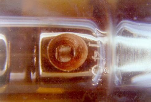
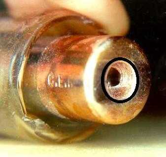
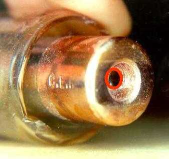
hands! LET'S ZOOM IN. What you see below is a close-up of the cathode, the negative end of the tube.
|
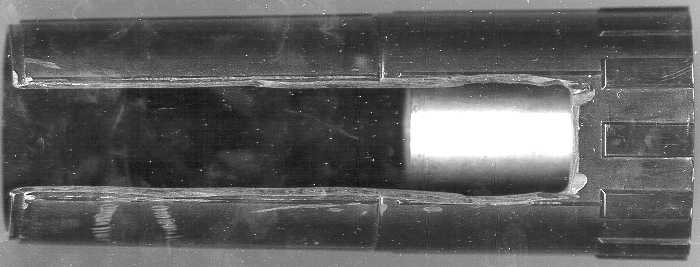
The glass of the envelope is only about 1 mm thick! The diameter of the stainless steel 'cup' is about 17 mm. The tungsten filament (that emits electrons when heated) is seen in the central rectangular recess (2 mm x 5 mm). The filament is very thin and has a length of about 5 mm. |
 It is time to
take a close look at the cathode and the anode.
It is time to
take a close look at the cathode and the anode.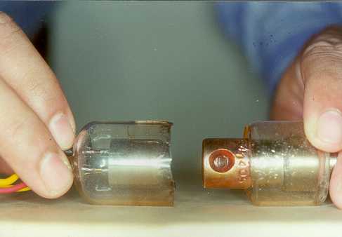

|
The electrons gather in the form of a cloud around the cathode, and get attracted towards the anode when high potential difference is applied through the cathode-anode (high-tension) circuit. |
|
|
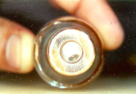
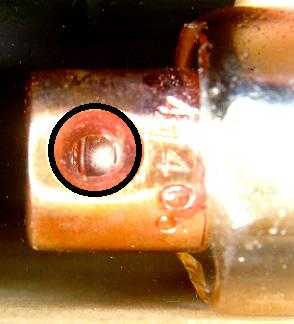
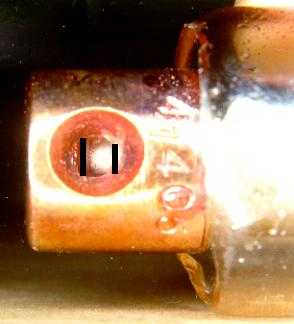
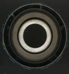
The anode is made up of a cylindrical copper block with two openings. The diameter of the copper block (as seen the picture on the left) is 17 mm. The opening (8 mm in diameter) in the front face of the copper anode allows entry of these electrons towards the tungsten focal spot. The opening (9 mm in diameter) in the side of the block allows exit of the x-ray beam out of the block. This opening actually acts as the so-called 'window'. (The text books describe the 'window' as a thinned portion of the glass envelope opposite the focal spot. No such thinned portion was evident in this x-ray tube.) Both these openings can be better appreciated in the pictures below: |
|
|
|
|
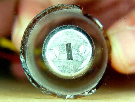
The black circle, this time, is outlining the 'window', which is actually a funnel-shaped opening. The outer diameter is 9 mm, the inner being 4 mm.The two black lines delineate the tungsten focal spot, which is angulated with respect to cathode ('principle of line focus').
|
|
|
The x-ray beam emergent from the tungsten focal spot passes through the window, the glass envelope, the surrounding oil and the plastic seal to reach the port (the 'inherent filtration'). The beam then encounters the aluminium filter (the 'added filtration') that removes the low-energy x-ray photons. The 'hardened' beam then has to be collimated to restrict it's shape and size. The tube head, that we dissected, had a cylindrical collimator.
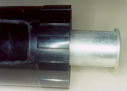
The picture on the above left shows the tube end of the collimator. The middle picture shows the metallic insert that collimates the x-ray beam. The picture on the right shows the plastic pointing device (partially cut open) to reveal the collimator.
|
|
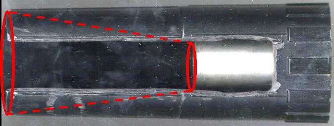 |
The beam that emerges at the outer end of the collimator diverges to give a size of 7-cms diameter at the skin. |
|
That was the story of our journey to the heart of a dental x-ray tube head. Thank you for joining us. You are welcome to use the pictures on these web pages for educational purposes. A line of acknowledgment ("pictures courtesy https://omr-cods.tripod.com/") would however be appreciated. Your comments on this case study and the rest of the web site can be entered in the guestbook. Please spare a few minutes of your time. |
Start the Story All Over Again.