|
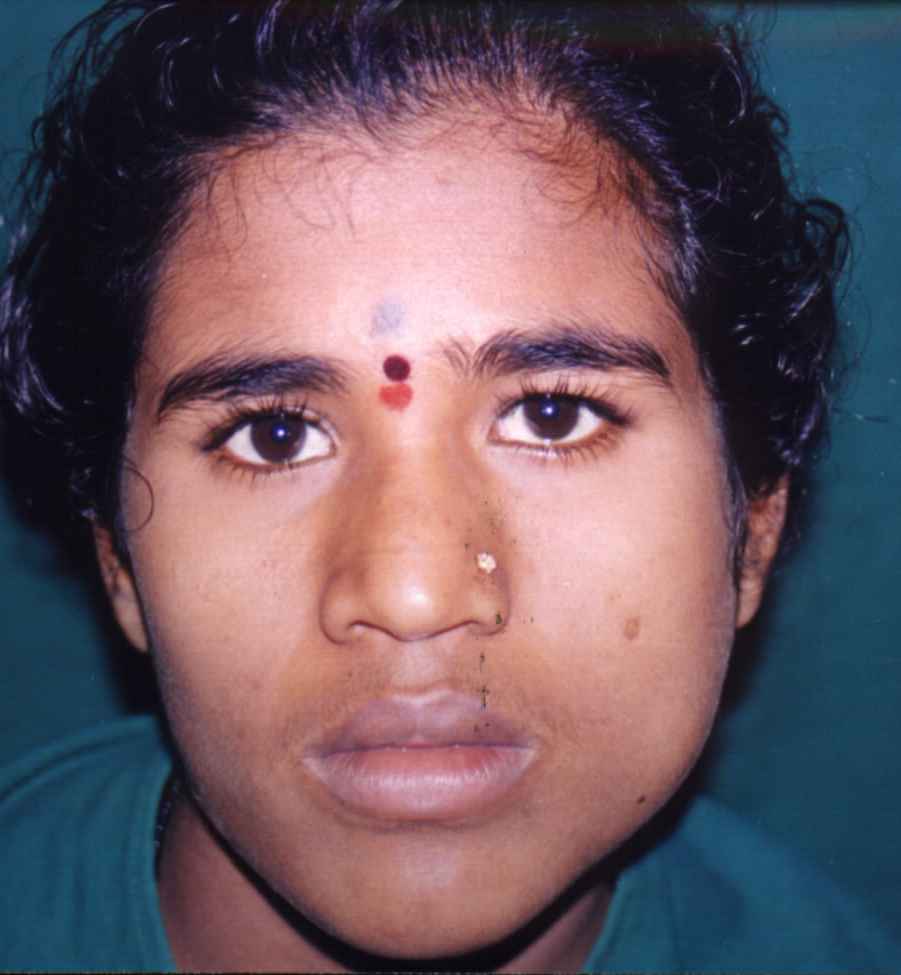
An 18-years old woman patient reported to the Dept. of Oral & Maxillo-facial Surgery with a complaint of pain and swelling in the left pre-auricular and mandibular angle region. The swelling was noticed about a month back to gradually increase to reach the present dimensions. Aching type of pain was experienced by the patient for the last 1 week. |
 |
 |
|
|
Patient
was in the 2nd trimester of pregnancy.
|
|||
| General examination: | |||
|
1. Conscious, co-operative patient with average built 2. No significant abnormality observed. |
|||
| Local examination: |
|
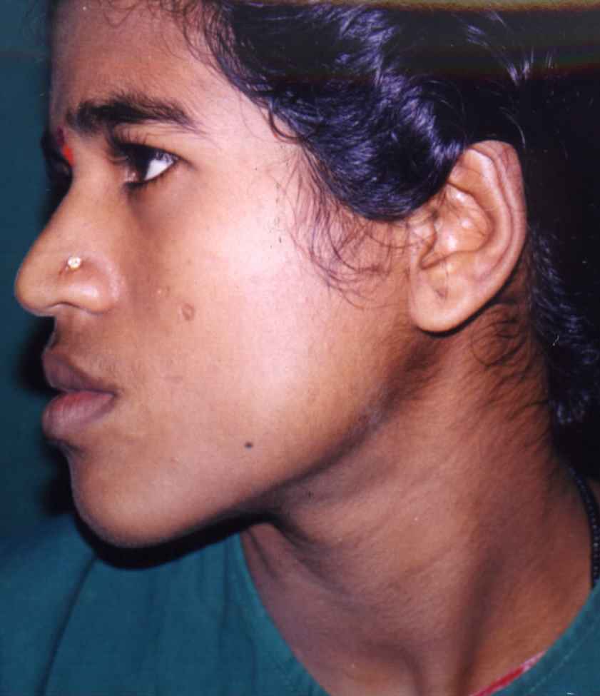
A diffuse
swelling was seen on the left side of the face extending from pre-auricular
region to the lower border of the mandible. The swelling extended from
the posterior border of the ramus to the anterior border of the masseter.
The overlying skin was normal in colour, temperature and texture, and
was pinchable. On palpation, it was hard in consistency. Egg shell crackling
and tenderness could be elicited along the lateral aspect of the swelling.
The regional lymph nodes were not palpable. |
|
|
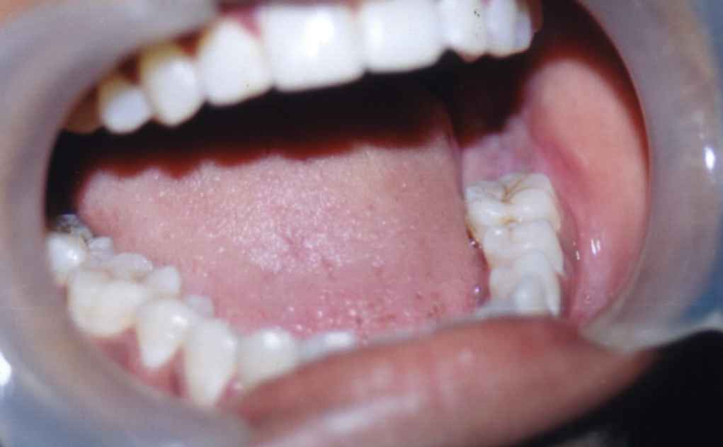
Intra-oral examination revealed the oral extension of the swelling that was diffuse in nature and soft in consistency. Tenderness was present. Teeth 37 and 38 were missing and tooth 36 exhibited mobility. The overlying mucosa was normal. Aspiration yielded straw color fluid with glistening cholesterol crystals. |
 |
| Radiographic examination: |
|
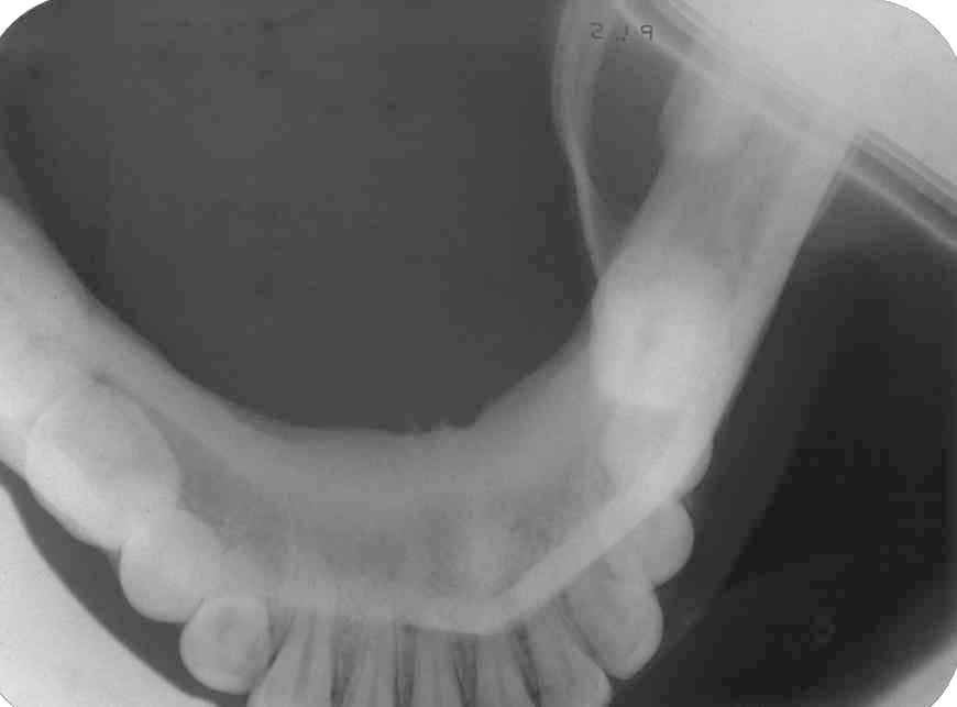
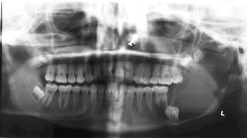
Intra-oral occlusal, panoramic, lateral oblique and PA-mandible projections were obtained. A large well-defined, unilocular radiolucency with hyperostotic border was seen. The radiolucency occupied the whole of the left ramus, including the coronoid process and extended upto the neck of the condyle. Anteriorly, the lesion extended upto the first molar. The anterior and posterior borders showed considerable thinning of the cortex. Scalloping of the margins was seen in the first molar region. The sigmoid notch was obliterated. The second molar was displaced to the lower border of the mandible, while the third molar was pushed into the coronoid process. The first molar roots showed some degree of resorption. |
|
|
 |
|
| Provisional diagnosis : | |||
|
The clinical behavior and the radiographic features suggested a dentigerous cyst, with a possibility of an ameloblastoma.
|
|
Clinical
Presentation
|
| Top |